

TriA SNOM
Scanning Near Field Optical Microscope
SNOM Working modes:
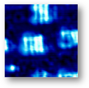
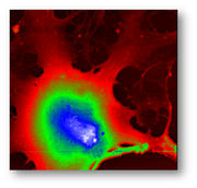
SNOM Topography: The image obtained is the result of signals acquisition in xyz axis that allows to measure detailed surface morphology and nanostructures on a nanometric scale.SNOM Optical Reflection: image is obtained with near-field light that interacts locally with a superficial layer of the sample, giving information on structures confined within 30-100 nm (depending on the tip aperture) below sample surface.
SNOM Optical Transmission: the image results from the light transmitted through the whole thickness of the sample. While in conventional optical microscope all the sample is illuminated, SNOM near-field light interacts only locally producing signals point by point. SNOM optical transmission images are comparable with conventional optical images but their lateral resolution is more than 10 times higher.
SNOM Optical Back-Reflection: image is created by the near-field light that is back-reflected into the fiber, after local interaction with the sample surface.
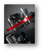
TriA SNOM Scanning system
Scanning stage with absolute positioning system and strain gauge sensors.Standard scanner technical data:
X-Y scan size:
- 100 x 100 μm (high voltage mode)
- 10 x 10 μm (low voltage mode)
- High voltage closed loop resolution: 2 nm
- High voltage open loop resolution: 0.2 nm
- Closed loop linearity: 0.1%
Z scan size:
- 10 μm (high voltage mode)
- 1 μm (low voltage mode)
- Resolution: 0.16 nm (high voltage mode), 0.02 nm (low voltage mode).
Based on specific demands other scanning ranges can be combined by the user in different configurations*.
Maximum sample size: it can accommodate samples with different geometries and sizes up to 30 mm diameter.
SPM Control System
It is composed by a digitally controlled analog feedback that combines the flexibility of computer controlled parameters with the high resolution and low noise of an analogue implementation. This detection scheme provides sub-nanometric vertical resolution in the images and all collected signals are distortions free.
The electronics supports STM, AFM and SNOM heads, performs different kinds of spectroscopy and can acquire several user-defined auxiliary channels.
Acquisition software
Software runs under Windows and is composed of a multi- window application to control the instrument and do the data acquisition. The software controls all the parameters of the instrument.
Tools & Accessories
TriA SNOM can be equipped with a large number of laser sources and filters
* Please contact your local distributor for specific configuration.
Characteristics and technical specifications subject to change